Overview of the BLA measurement
The spectrometer for which this software was developed uses a bent Laue analyzer of the sort first described by Zhong et al. The analyzer is in the form of a log-spiral. A sheet of silicon crystal (most of the examples in this document use a Si (220) crystal) is pressed against this log-spiral surface.
The log-spiral is the shape that preserves the divergence of the signal fluorescing from a sample as the light is diffracted from the planes of the crystal. This, then, serves to spatially separate the light diffracted by the crystal from the light transmitted through the crystal, as seen in this schematic:
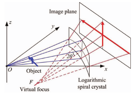
Schematic of the log-spiral bent Laue analyzer, taken from figure 1 in Wu et al.
If an area detector is placed far enough away from the analyzer, only the light diffracted by the crystal will be incident upon the face of the detector.
- Yufen Wu, Shali Xiao, Jian Lu, Lifeng Liu, Qingguo Yang, and Xianbin Huang. Research on a logarithmically bent laue crystal analyzer for x-ray monochromatic backlight imaging. Review of Scientific Instruments, 2013. doi:10.1063/1.4815549.
- Z. Zhong, L. D. Chapman, B. A. Bunker, G. B. Bunker, R. Fischetti, and C. U. Segre. A bent Laue analyzer for fluorescence EXAFS detection. Journal of Synchrotron Radiation, 6(3):212–214, May 1999. doi:10.1107/S0909049599002022.
In practice, it is useful to use some kind of collimator, such as Soller slits, to suppress background on the detector from light scattering from other parts of the experiment.
Here is a photograph of the spectrometer as it was used in February 2016.
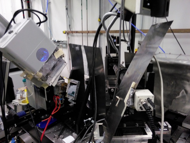
The shrouded box at the far right of the photo contains the Kirkpatrick-Baez focusing mirrors used to deliver a 10s-of-microns sized small spot to the sample. The shielding is in place to keep light from scattering off the mirror and into the detector.
The sample is mounted on an XY stage with an optical camera observing the sample.
The analyzer is the shiny object just below and to the left of the center of the center of the photo. The shiny part is the Si (220) crystal. The aluminum frame around the Si is made in the log-spiral shape.
The sheets of metal between the sample and the analyzer are for shielding light that might scatter from the region of the sample in the direction of the detector.
Suspended at the appropriate angle to intercept the light diffracting from the analyzer is a Pilatus area detector. Between the analyzer and the Pilatus are a set of Soller slits.
Several parts of the spectrometer are actuated. The sample can be moved in the incident beam. The optical camera is used to bring the surface of the sample to the same spot after a sample change.
The analyzer can rotate about it's diffraction axis and can move closer to or farther from the sample. Together, these motions bring the analyzer into focus with the sample at a given energy.
The Pilatus is rotated to twice the angle of the analyzer crystal so that the diffracted light enters its window.
In the following sections, examples of HERFD, XES, and RIXS measurements using the instrument are shown.
Xray::BLA and METIS are copyright © 2011-2014, 2016 Bruce Ravel and Jeremy Kropf – This document is copyright © 2016 Bruce Ravel
This document is licensed under The Creative Commons Attribution-ShareAlike License.
If this software and its documentation are useful to you, please consider supporting The Creative Commons.